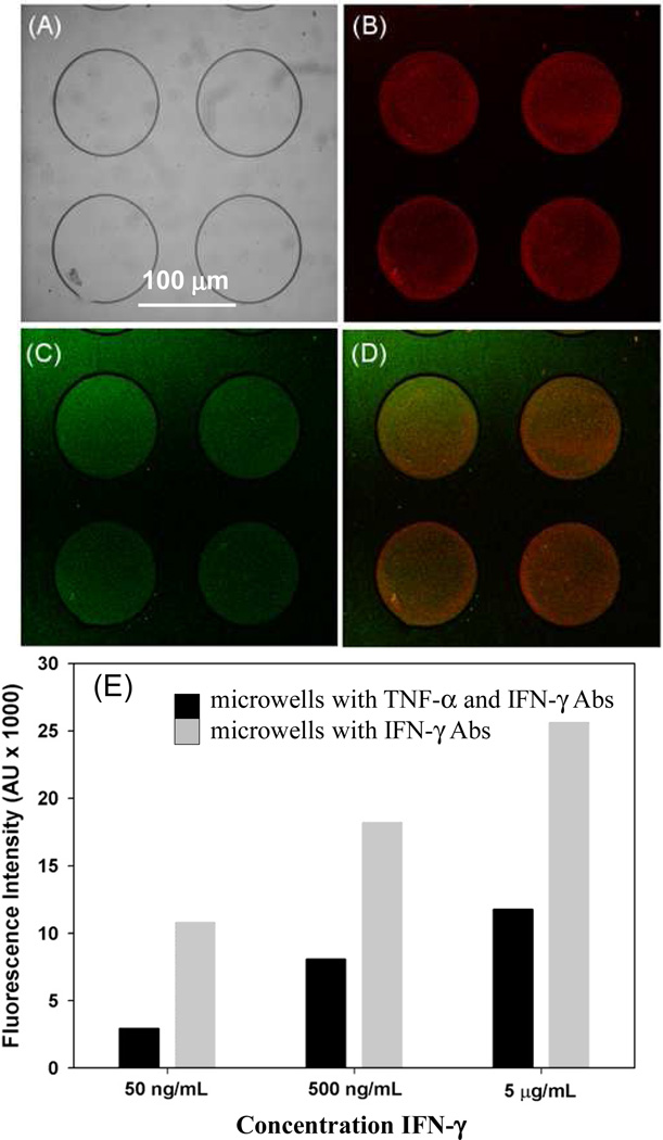
Figure 6.
Detection of multiple cytokines inside hydrogel microwells. PEG hydrogel microwells were fabricated on a mixed silane layer, functionalized with neutravidin and then incubated with a 1:1 mixture of anti-IFN-γ and anti-TNF-α Abs. (A) Brightfield image of hydrogel microwells. (B–D) These microwells were simultaneously challenged with 500 ng/mL IFN-γ and TNF-α, subsequently microwells were incubated in a mixture of anti-IFN-γ-PE and anti-TNF-FITC. Images show both red and green fluorescence inside the microwells demonstrating that the two cytokines could be detected simultaneously. (E) Characterizing sensitivity of cytokine immunoassay as a function of Ab immobilization. Responses to 500 ng/mL (30 nM) IFN-γ were compared for microwells modified with anti-IFN-γ as well as microwells containing anti-IFN-γ/anti-TNF-α combination.

