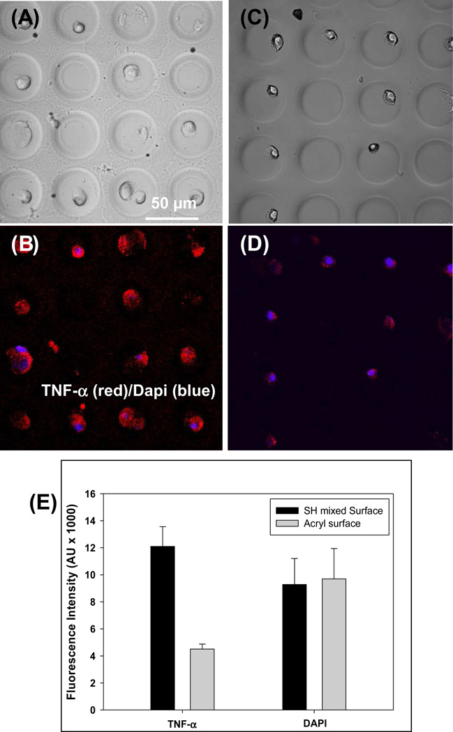
Figure 8.
Detection of TNF-α release from individual macrophages. PEG hydrogel microwells (30 µm diameter) were precoated with anti-TNF-α Abs and incubated with cells. (A–D) Imaging of macrophages in hydrogel microwells. Brightfield images of cells captured in microwells constructed on mixed silane (A) and mono-functional silane (C). Immunofluorescent staining for TNF-α production in activated macrophages (B and D). Cells were mitogenically stimulated for 3 h and then stained with anti-TNF-α-biotin followed by streptavidin-Alexa546. Cell nuclei were stained with Dapi (blue color). (E) Characterization of TNF-α fluorescence in microwells created on bi-functional or mono-functional silanes. Microwells with covalently immobilized avidin-biotin-Ab construct had 3 fold higher fluorescence signal compared to sensing microwells created on monfunctional silanes. Dapi intensity was the same for cells captured in both types of microwells. `The signal represents an average fluorescence in 20 microwells.

