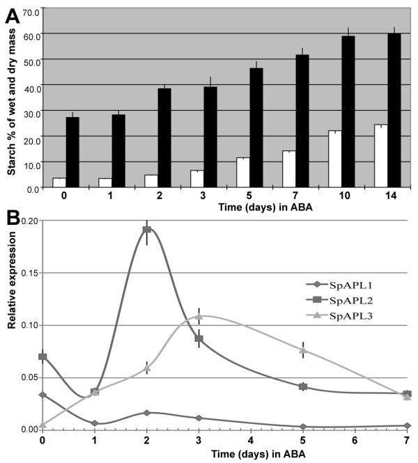
Figure 2.

Starch accumulation and expression of the APL genes during turion development. a) White bars stands for wet tissue and black bars for dry tissue. Y-axis shows starch content (mg) for every 100 mg wet tissue or dry tissue. b) qPCR was used to quantify expression of APLs based on RNA from 0, 1, 2, 3, 5, 7 days of ABA treatment. Standard error was shown by vertical bar. Sample collections for starch analysis and APLs expression quantification were listed in Table 1.
