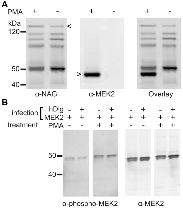
Figure 3.
hDlg interacts with activated MEK2. A GST-hDlg fusion protein and MEK2 were co-expressed in insect cells. Lysates from PMA-treated (+) and untreated (-) cells were incubated with GSH-agarose and the content of the bound fractions was submitted to SDS-PAGE and transferred onto nitrocellulose. The membrane was probed with anti-hDlg (α-NAG) and anti-MEK2 (α-MEK2) antibodies. The membrane was scanned with a dual laser Odyssey system to co-detect hDlg (left panel, A) and MEK2 (middle panel, A). The right panel in A shows the overlay of the two scans. Full-length hDlg and full-length MEK2 are identified by arrow heads. The multiple bands detected by anti-hDlg antibodies correspond to breakdown products consistently observed when hDlg is overexpressed in insect cells. Panel B shows immunoblots of lysates from PMA-treated (+) and untreated (-) cells that were infected with baculovirus for the expression of MEK2 alone or in combination with baculovirus driving the expression of hDlg and probed with anti-MEK2 (α-MEK2, left) and anti-phospho-MEK1/2 (α-phospho-MEK2, right).

