
Figure 8.
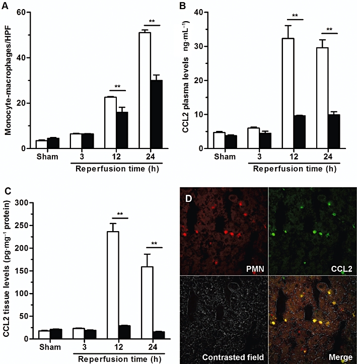
DF 2156 inhibited monocyte/macrophage infiltration and CCL2 production originating from infiltrating PMN into post-ischaemic rat liver. Reperfusion injury was induced by 1 h ischaemia of the liver followed by 3, 12 or 24 h reperfusion. Ischaemic rats were treated i.v. with DF 2156A (15 mg·kg−1; solid columns) or vehicle (open columns) 15 min before reperfusion (six animals per experimental group). (A) Detection of monocytes/macrophages after 3, 12 or 24 h of reperfusion was performed using the mouse monoclonal antibody (MCA 341R) against ectodysplasin, the rat homologue of human CD68. Monocytes/macrophages present in sinusoids and extravasated into the parenchymal tissue were counted in 20 non-consecutive, randomly chosen 400× histological HPF. CCL2 concentration was determined in plasma (B) or hepatic tissue (C) after 3, 12 or 24 h of reperfusion using rat CCL2 immunoassay kit. Data are mean ± SEM from one experiment representative of three performed. *P < 0.05; **P < 0.01 versus respective vehicle-treated group by Student's t-test. (D) Confocal analysis of CCL2 expression was determined in liver frozen and fixed sections from vehicle-treated rats 24 h after reperfusion. Slides were incubated with a mix of primary antibodies (mouse anti-rat PMN and hamster anti-mouse CCL2) overnight at 4°C. Sections were then rinsed, incubated with a mix of secondary antibodies (goat anti-mouse IgM Alexa Fluor 546 and goat anti-hamster FITC) for 1 h at room temperature, mounted with glycerol and analysed with a 510 Laser Scanning Microscope. Infiltrated PMNs (red stained) and CCL2 expression (green stained) showed an almost total colocalization (merge image).