Figure 5.

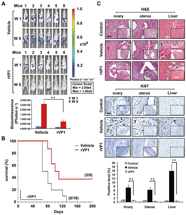
rVP1 attenuates tumour growth in an intraperitoneal SKOV3-xenograft mouse model. Intraperitoneal injection with vehicle or 15 mg·kg−1 rVP1 was started at 6 days after the SKOV3-GL inoculation. (A) Bioluminescent images showing the presence of SKOV3-GL cells in vivo. Bar graphs represent photon intensity (means ± SEM). (B) Comparison of survival rates between vehicle- and rVP1-treated SKOV3-xenograft mice. Vehicle treatment t50 = 74.5 day, n = 10; rVP1 treatment t50 = 122.5 day, n = 8; P < 0.05. (C) Ovaries, uterus and livers were collected from mice and processed for haematoxylin and eosin [H&E] staining and immunohistochemical analysis with anti-human Ki67 antibodies (indicated by red arrow). Tissue slides were scanned and analysed using ImageScope 9.1 software. Data represent means ± SD of eight fixed fields within the tumour section. Scale bars in panel C, 100 µm. **P≤ 0.005.
