
Figure 6.
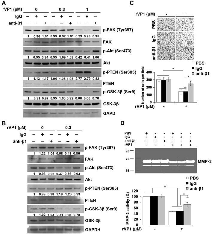
rVP1 suppresses invasive capacity and MMP-2 activity of SKOV3ip.1 cells through integrin β1-Akt signalling. (A) SKOV3ip.1 cells were isolated from the ascites of intraperitoneal SKOV3-xenograft mice at 60 days after the implantation. SKOV3ip.1 cells were pretreated with or without control IgG or anti-integrin β1 antibody (2 µg·mL−1) for 1 h followed by 0.3 or 1 µM of rVP1 treatment for 30 min as indicated. After incubation, lysates were analysed by Western blotting for FAK, p-FAK, Akt, p-Akt, GSK-3β, p-GSK-3β, PTEN and p-PTEN. (B) SKOV3ip.1 cells were treated with or without 0.3 µM rVP1 for 24 h at 37°C in serum-free medium. After incubation, lysates were analysed by Western blotting. Blots are representative of three independent experiments. (C, D) SKOV3ip.1 cells were pretreated with control IgG or anti-integrin β1 antibodies (2 µg·mL−1) for 30 min followed by 0.3 µM rVP1 treatment for 24 h in serum-free medium. The invasive capacity of the cells was determined by Matrigel invasion assay. The MMP-2 enzyme activity in the cell culture medium was analysed by a gelatinolytic zymography assay. Data represent means ± SD of three independent experiments; *P < 0.01.