
Figure 6.
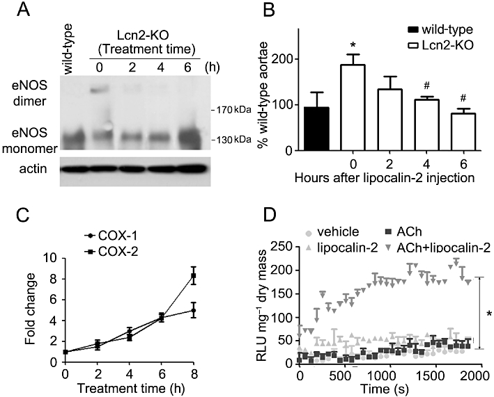
Lipocalin-2 was administered into Lcn2-KO mice as in Figure 5. Aortae and carotid arteries were collected at different time points for Western blotting and quantitative PCR to analyse the dimers and monomers of eNOS (A), NO production (B) and COX expressions (C), respectively. The % NO of wild-type aortae was presented from the averages of four experiments. *P < 0.05 versus wild-type mice; #P < 0.05 versus Lcn2-KO mice at time zero; n = 5. The quantitative PCR results are expressed as fold changes versus time zero. Carotid artery collected at 6 h after protein injection was subjected to lucigenin-enhanced chemiluminesence assay as in Figure 4. The results are expressed as relative luminescence unit (RLU) normalized against dried tissue masses (D). Note that lipocalin-2 significantly enhanced NADPH oxidase activity stimulated by ACh (10−6 M). *P < 0.05 ACh + lipocalin-2 versus other groups; n = 3.