
Figure 7.
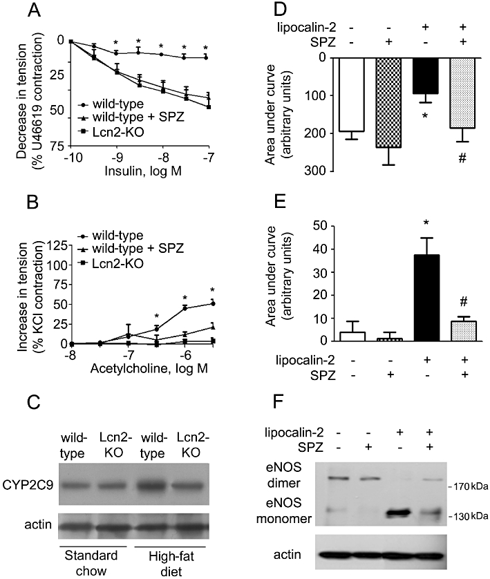
Left: SPZ or vehicle was administered to wild-type mice (under high-fat diet) by i.p. injection. At the end of the treatment period (3 weeks), the aortae and carotid arteries were collected for evaluation of endothelium-dependent relaxations to insulin (A) and endothelium-dependent contractions to ACh (B), respectively. The amount of CYP2C9 protein in aortae of wild-type and Lcn2-KO mice was monitored by Western blotting (C). *P < 0.05 versus the other two groups; n = 5. Right: same treatment was performed in Lcn2-KO mice fed a high-fat diet. At the end of the treatment period (3 weeks), mice were injected with lipocalin-2 protein as described in Figure 5. Six hours after injection, the aortae and carotid arteries were collected for evaluation of endothelium-dependent relaxations to insulin (D) and endothelium-dependent contractions to ACh (E), respectively. Data are presented as areas under curve. Western blotting was performed for analysing the dimers and monomers of eNOS in aortae of these mice (F). *P < 0.05 versus vehicle-treated mice; #P < 0.05 versus lipocalin-2-treated mice; n = 5.