Figure 2.
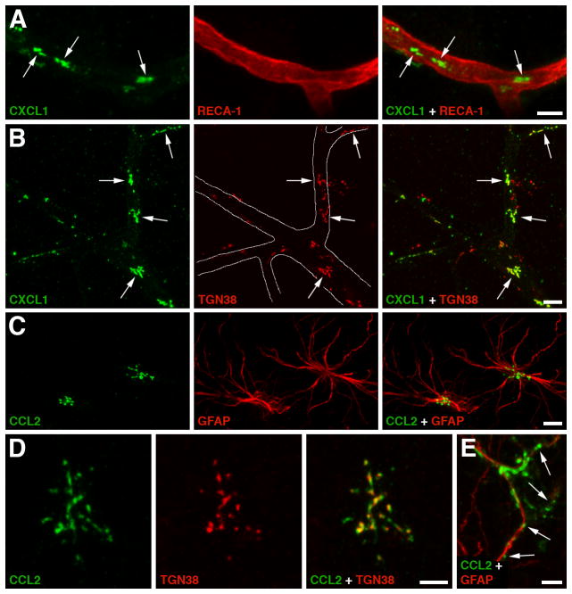
Production of CXC and CC chemokines by the cerebrovascular endothelium and astrocytes in a rat model of traumatic brain injury (TBI). A The immunoreactive product for neutrophil chemoattractant CXCL1 (arrows) associated with a microvessel in the traumatized brain parenchyma at 6 hours post-TBI. Double immunostaining with anti-CXCL1 antibody and an antibody to RECA-1, a marker for vascular endothelium, is shown. Similar results were obtained for CXCL2 and CCL2, a major chemoattractant for monocytes. This distinct pattern of immunopositive staining for CXC and CC chemokines was not associated with pericytes, perivascular macrophages, or smooth muscle cells, suggesting that these chemokines are produced by the brain endothelium. B Double immunostaining with anti-CXCL1 antibody and an antibody to TGN38, a Golgi marker, demonstrates that the CXCL1-immunoreactive product in cerebral microvessels (outlined) is predominantly associated with the Golgi complex (arrows). The localization of CXC and CC chemokines to the Golgi complex is consistent with increased synthesis of these secreted proteins occurring after injury. C Post-traumatic expression of CCL2 in astrocytes. Double immunostaining with anti-CCL2 antibody and an antibody to glial fibrillary acidic protein (GFAP), an astrocyte marker, is shown. Neither CXCL1 nor CXCL2 were expressed in astrocytes after injury. D CCL2 expressed in a cortical astrocyte at 6 hours post-TBI co-localizes with the Golgi complex. E Astrocytic expression of CCL2 at 24 hours after TBI. Note that at this time after injury, the CCL2-immunopositive product is predominantly localized extracellularly along astrocyte processes (arrows). This pattern of CCL2-immunopositive staining likely reflects the binding of this chemokine to heparan sulfate proteoglycans expressed on the surface of astrocytic cells and/or within the extracellular matrix. Scale bars: panels A–C, 10 μm; panels D, E 5 μm. Reprinted with permission from [178].

