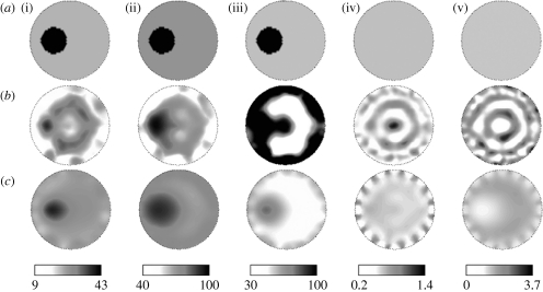
Figure 3.
Comparison of images obtained by applying the spectrally constrained direct chromophore reconstruction and the conventional technique of separate wavelength optical properties recovery on measurements from a gelatin phantom with a 25 mm inclusion. The gelatin phantom contained whole blood and TiO2 for scattering and the inclusion was filled with 4% pig blood and 0.75% Intralipid in buffered saline. (a) The expected images are shown for (i) [HbT] (μM), (ii) oxygen saturation S tO2(%), (iii) water H2O(%), (iv) scattering amplitude a and (v) scattering power b, along with (b) conventional technique images and (c) spectral method images.

