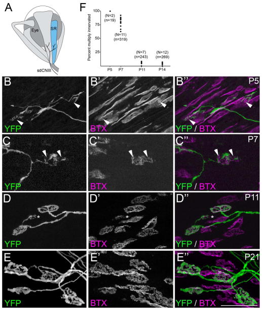
Fig. 1. Development of motor nerve terminals and AChR clusters at ‘en plaque’-type synapses in SR muscle.
A. Schematic representation of SR muscle (in blue) and its innervation by the superior division of the oculomotor nerve (sdCNIII). B–E. YFP-expressing motor nerve terminals (green) were imaged in P5 (B), P7 (C), P11 (D) and P21 (E) SR muscles isolated from thy1:yfp line H mice. AChR clusters were simultaneously visualized by labeling with fluorescently-conjugated bungarotoxin (BTX; magenta). At early ages (B,C) YFP-containing motor nerve terminals failed to innervate the entire postsynaptic membrane, suggesting that synaptic sites remained multiply innervated at these ages. Arrowheads highlight AChR-rich regions of the postsynaptic membrane that were unoccupied by YFP-labeled nerve terminals. By P11 (D), few synaptic sites contacted by YFP-labeled axons appeared multiply innervated. Asterisk in D, highlights an ‘en grappe’-type AChR cluster contacted by the same motor axon that contacted 2 ‘en plaque’-type NMJs in this field of view. Scale bar = 50 μm. F. Numbers of multiply innervated NMJs were quantified at P5, P7, P11, and P14 from thy1:yfp line H SR muscles. Each black dot represents the percentage of YFP-labeled NMJs that were multiply innervated from a single muscle. N = number of muscles analyzed per age. n = total of number of NMJs analyzed per age.

