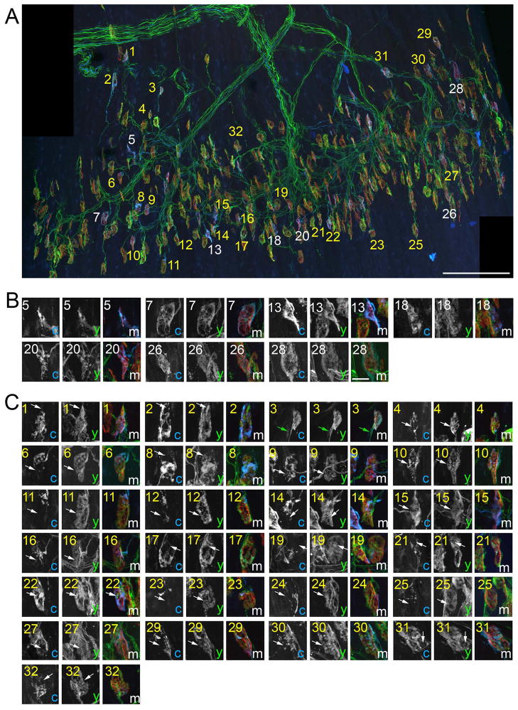
Fig 5. Multiple nerve terminals were observed at most synaptic sites in P10 LPS muscles.
To conclusively determine synaptic sites in P10 LPS muscle were contacted by multiple axons, NMJs were imaged in thy1:yfp line 16; thy1:cfp line23 mice in which all motor axons were labeled with YFP and only a few were also labeled with CFP. A. A montage of high-resolution images encompassing the entire central end-plate band. AChR clusters were labeled with BTX (red). 32 NMJs contacted by a CFP-labeled axon were labeled with numbers: white numbers depict singly innervated NMJs; yellow numbers depict synapses contacted by both CFP-labeled axons and YFP-labeled (CFP-non-labeled) axons. B. High magnification images of singly innervated NMJs from (A). C. High magnification images of NMJs that are contacted by both a CFP-expressing axon and supernumerary YFP-expressing axons. Arrows in C highlight domains of these NMJs that are innervated by YFP-labeled (CFP-non-labeled) nerve terminals. In some cases although CFP-labeled terminals appeared to fully occupy a postsynaptic site, YFP-labeled (CFP-non-labeled) axons were still observed co-innervating these site (see green arrows in image number 3 of C). c = CFP; y = YFP; m = merge. Scale bar in A = 200 μm, in B = 10 μm for B,C.

