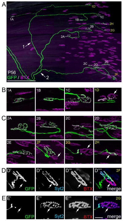
Fig 8. Reconstruction of two LPS motor units revealed multiply innervated synaptic sites persisted at P56.
A. Confocal reconstruction of two motor axons innervating the same P56 LPS muscle from a thy1:gfp line S mouse. Each axon is labeled (with arrows and either a 1 or 2) as they course toward the end-plate band. Synaptic sites contacted by each axon are labeled with numbers that correspond to the axon innervating them and a letter. Sites labeled in white appear singly innervated; sites labeled in yellow synapses appear multiply innervated. B. High magnification images of synaptic sites in motor unit 1. Arrow indicates AChR-rich regions of a synaptic site not fully innervated by the GFP-labeled axon. C. High magnification images of synaptic sites in motor unit 2. Arrows indicates AChR-rich regions of synaptic sites not fully innervated by the GFP-labeled axon. D,E. After imaging, the LPS muscle was immunostained for synaptotagmin 2 (Syt2), a component of the presynaptic machinery within all motor nerve terminals. Immuno-labeling for Syt2 (blue) revealed that portions of the synaptic sites not occupied by the GFP-labeled were occupied by other axons (see arrows). Scale bar in A = 250 μm and in E = 10 μm for B–E.

