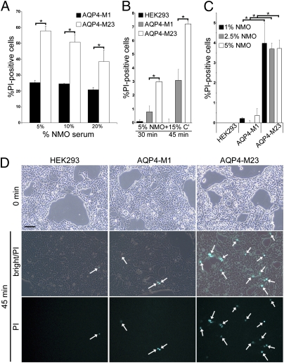
Fig. 4.
M23 interaction with NMO-IgG activates complement more effectively than M1. Simultaneous addition of complement and heat-inactivated NMO serum caused rapid lysis of HEK293 cells expressing M1 or M23. Therefore, for flow cytometric analysis cells were exposed sequentially to serum (30 min at 22 °C) and complement (45 min at 37 °C) (4). Lesioning (PI permeability) was negligible with heat-inactivated complement (values subtracted). Bars represent SEMs of three or more experiments. (A) Cells exposed to graded concentrations of NMO serum and 20% complement. Membrane lesioning was maximal with 5% NMO serum [M1 compared with M23; *P < 0.001 (5%); *P < 0.001 (10%); *P = 0.013 (20%)]. (B) To circumvent M1 internalization by NMO-IgG at 37 °C, we simultaneously added 5% serum and complement at suboptimal concentration (15%). M23 cells showed consistently greater vulnerability to lesioning [*P = 0.031 (30 min); *P = 0.013 (45 min)]. (C) With complement limited to 10% concentration for 45 min, its activation by M23 cells was consistently fourfold more than by M1 cells at all tested NMO serum concentrations [*P = 0.05 (1%); *P = 0.001 (2.5%); *P = 0.005 (5%)]. (D) Phase-contrast microscopy appearance of PI permeable cells (cyan; arrows) under conditions shown in B (nontransfected and transfected with M1 or M23). (Scale bar, 200 μm.)

