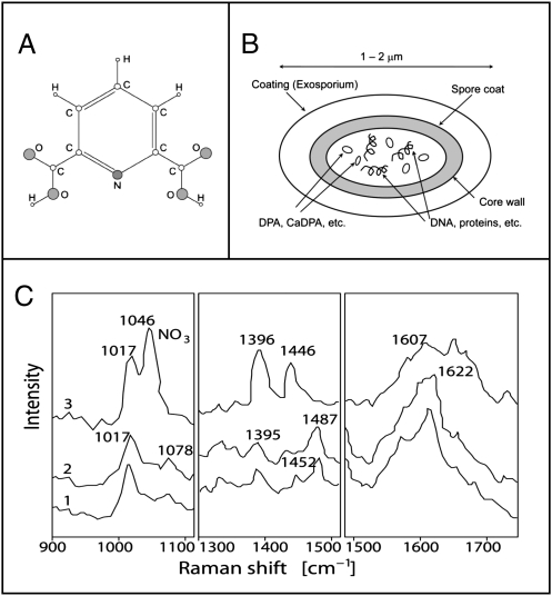
Fig. 1.
(A) Chemical structure of DPA. (B) Sketch of a bacterial endospore that indicates that the DPA and its salts (e.g., Ca-DPA) are contained in the core. (C) UV resonance Raman spectra of spores of Bacillus megaterium (trace 1), spores of Bacillus cereus (trace 2), and calcium dipicolinate (trace 3) in three spectral regions. All samples are excited at 242 nm. Modified from ref. 1.

