Fig 6.
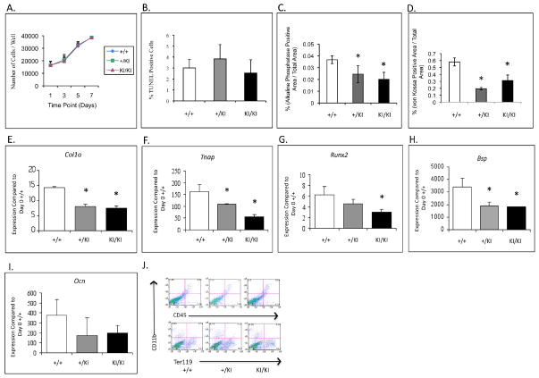
Calvarial osteoblast culture assays. A) and B) Growth curve and apoptosis assays. No significant difference in cell growth and apoptosis between control (Sh3bp2+/+) and mutant cultures (Sh3bp2+/KI Sh3bp2KI/KI). C) and D) Differentiation and mineralization assays. Significant decrease in alkaline phosphatase and von Kossa stained areas in Sh3bp2KI/KI cultures. E–I) QPCR analysis. Gene expression of osteoblast differentiation markers is significantly decreased in Sh3bp2KI/KI cultures as compared to control (Sh3bp2+/+) cultures and J) FACS analysis of freshly isolated calvarial osteoblasts after hematopoietic cell depletion. There is no significant difference in levels of CD45, CD11b and Ter119 positive cells between Sh3bp2KI/KI and Sh3bp2+/+ calvarial osteoblasts.

