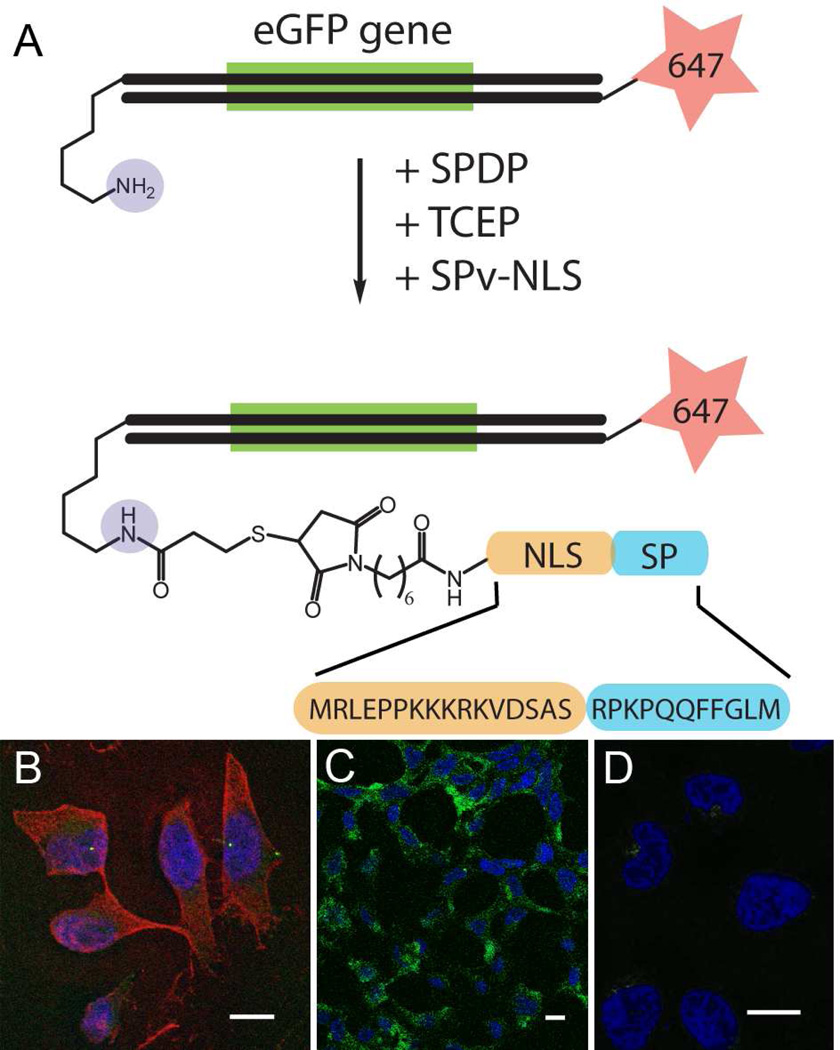
Fig. 2.
Delivery of functionally-expressed DNA fragment. A. A DNA fragment coding for GFP was generated by PCR with the introduction of Alexa-647 fluorophore (red) and a free amine (blue). SPDP was used to conjugate the DNA fragment to SPv-NLS, which contains an N-terminal maleimde, followed by a nuclear localization sequence (orange) and the SP sequence. B. Hela cells transfected with the NK1R show internalization of the DNA conjugate (red) with both cytoplasmic and nuclear localization after 2 hrs of treatment with 200 pM DNA-SPv-NLS conjugate, and C. GFP expression (green) after 48 hrs of treatment. D. Hela cells treated with DNA fragment not conjugated to SP. (Scale bar = 10 µm).

