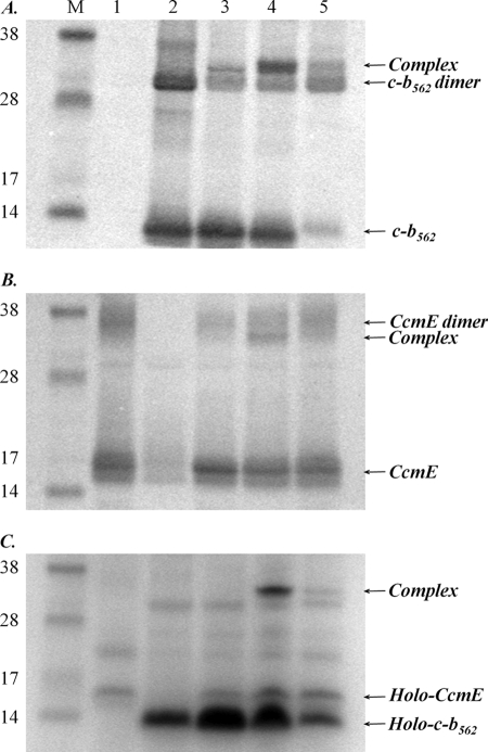
FIGURE 2.
Detection of a complex between CcmE, heme, and cytochrome c-b562. A, Western blot of cell membranes using a cytochrome b562 antibody. B, Western blot of cell membranes using a CcmE antibody. C, SDS-PAGE of cell membranes stained for proteins containing covalently bound heme. For each panel, the lane order is: M, molecular mass markers (as indicated, in kDa); lanes 1, cells expressing the Ccm system from pEC86; 2, cells expressing cytochrome c-b562 R98C/Y101C; 3, cells expressing the Ccm system from pEC86 and cytochrome c-b562 R98C/Y101C; 4, cells expressing the Ccm system from pEC86 and cytochrome c-b562 R98C; 5, cells expressing the Ccm system from pEC86 and cytochrome c-b562 Y101C. The position of the CcmE-heme-cytochrome c-b562 complex, which runs just above a cytochrome c-b562 dimer and just below the CcmE dimer, is indicated. An equal amount of total protein was loaded in each lane.

