FIGURE 5.
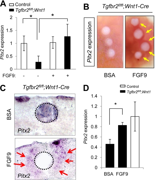
Exogenous FGF9 rescues Pitx2 expression in Tgfbr2fl/fl;Wnt1-Cre mice during palatal development. A. Quantitative RT-PCR analysis of Pitx2 in primary MEPM cells of Tgfbr2fl/fl control (open columns) and Tgfbr2fl/fl;Wnt1-Cre (closed columns) mice after treatment (+) or no treatment (−) with FGF9 protein. *, p < 0.05. B, whole mount in situ hybridization after implantation of FGF9 or BSA control beads in the palate of Tgfbr2fl/fl;Wnt1-Cre mice. The arrows indicate the increased expression of Pitx2 around FGF9 beads. C, section in situ hybridization for Pitx2 after implantation of FGF9 or BSA control beads in the palate of the Tgfbr2fl/fl;Wnt1-Cre mice. The arrows indicate the increased expression of Pitx2 around FGF9 beads. Dotted lines, perimeter of the beads. D, quantitative RT-PCR analysis of Pitx2 in the mesenchyme of Tgfbr2fl/fl;Wnt1-Cre (closed columns) and Tgfbr2fl/fl control (open column) mice after FGF9 or BSA treatment. *, p < 0.05. Error bars, S.D.

