FIGURE 7.
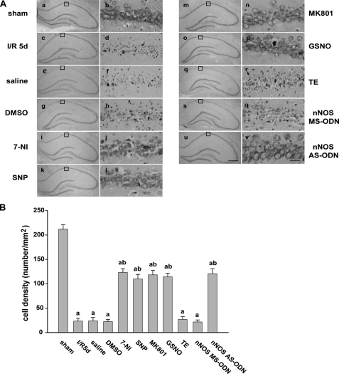
Effect of NOS inhibitors and exogenous NO and NMDAR antagonists on the survival of CA1 pyramidal neurons. A, cresyl violet staining was performed on sections from the hippocampi of sham-operated rats (panels a and b) and of rats subjected to 5 days of reperfusion after global ischemia (panels c and d), and the administration of saline (panels e and f), DMSO (panels g and h), 7-NI (panels i and j), SNP (panels k and l), MK801 (panels m and n), GSNO (panels o and p), TE (panels q and r), MS-nNOS (panels s and t), and AS-nNOS (panels u and v) before or after ischemia. Cresyl violet staining data were obtained from six independent animals, and a typical experiment is presented. B, cell density was expressed as the number of cells per 1-mm length of the CA1 pyramidal cells counted under a light microscope. Data are the mean ± S.D. (n = 6). Scale bars, 300 μm (panels a, c, e, g, i, k, m, o, q, s, and u); 30 μm (panels b, d, f, h, j, l, n, p, r, t, and v). a, p < 0.05 versus the sham groups; b, p < 0.05 versus the saline groups.

