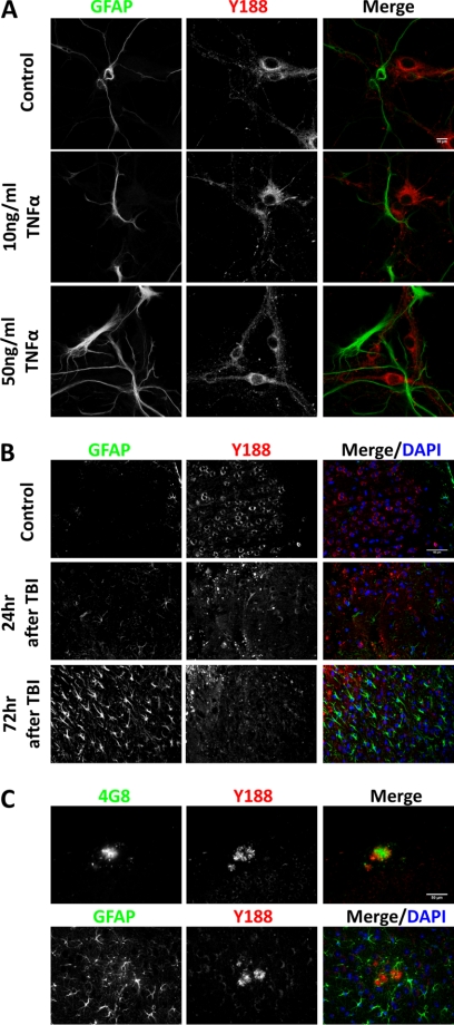
FIGURE 3.
APP expression is not induced in activated astrocytes in vitro and in vivo. A, 12 DIV primary hippocampal cultures were treated with PBS (Control), 10 ng/ml TNFα, and 50 ng/ml TNFα followed by immunocytochemical staining for APP. Blue, DAPI; Green, GFAP; Red, Y188. Scale bar, 10 μm. B, controlled cortical impact-induced TBI was performed on adult mouse brain. APP immunostaining was carried out in uninjured controls (Control), 24 or 72 h after TBI. Blue, DAPI; Green, GFAP; Red, Y188. Scale bar, 50 μm. C, immunostaining of 20 month-old APP/hAβ/PS1 brains. Upper panel, APP detected by Y188 (red) was found around 4G8-positive Aβ plaques (green). Lower panel, GFAP-positive reactive astrocytes (green) do not express APP (red). Scale bar, 50 μm.

