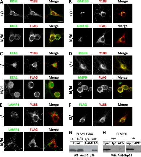
FIGURE 4.
Subcellular localization of FL-APP and APPsβ. A–E, 14 DIV hippocampal neurons cultured from wild-type (+/+) or homozygous APPsβ knock-in (ki/ki) mice were co-stained with APP (red) and organelle markers (green). A, ER (anti-KDEL); B, Golgi (GM130); C, early endosome (EEA1); D, late endosome (M6PR); E, lysosome (LAMP1). APP and APPsβ were recognized by Y188 or anti-FLAG antibody, respectively. F, co-staining of FL-APP and APPsβ in APPsβ ki/+ neurons. Green, FLAG; Red, Y188. G, interaction between Grp78 and the APP soluble domain. Wild-type (+/+) or homozygous APPsβ knock-in (ki/ki) brain lysates were immunoprecipitated (IP) using the anti-FLAG antibody conjugated to magnetic beads followed by elution with 3×FLAG peptide and Western blotting (WB) using an antibody against Grp78. Input, total lysates. H, interaction between Grp78 and FL-APP. Wild-type (+/+) or APP-null (−/−) brain lysates were immunoprecipitated using the APPc antibody or rabbit IgG plus protein-G beads followed by Western blotting using an antibody against Grp78.

