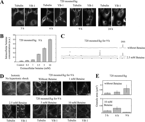
FIGURE 5.
The accumulation of compatible osmolytes after hypertonic exposure promotes SG disassembly. A, shown is time-lapse immunolabeling of NRK cells labeled with anti-YB1 and anti-tubulin after hypertonic shock. Up to 9 h of hypertonic exposure, SG size grew with time, and MT bundles were thicker. Between 9 and 24 h, SGs and MT bundles dissociated in the few cells that survived. Scale bar, 15 μm. B, we analyzed by NMR the intracellular betaine accumulation in NRK cells after a hypertonic shock with various extracellular concentrations of betaine for 9 h. The results show that betaine transport in NRK cells under hypertonicity is accelerated in the presence of extracellular betaine. Results are the means ± S.D., and 1 a.u. corresponds to 72 fmol of betaine per NRK cell (see “Materials and Methods”). C, two typical NMR spectra were obtained as described in B. In the presence of increasing betaine concentration, the area of cellular betaine peaks (see the asterisks) significantly increases. D, NRK cells were exposed for 9 h to constant hypertonicity in the presence of varying concentrations of betaine. Betaine, above 2.5 mm, promotes the disassemblies of SGs and MT bundles. SGs and MTs were detected with anti-YB1 and anti-tubulin immunostaining, respectively. Scale bar, 15 μm. E, shown are statistical measurements of the mean SG area obtained from the analysis of NRK immunostained with anti-YB1 after the indicated treatment. Although SG size increased between 6 and 9 h for hypertonic-stressed cells in the absence of betaine, a net decrease in SG size was detected in the presence of 10 mm betaine. Results are the means ± S.D.

