FIGURE 2.
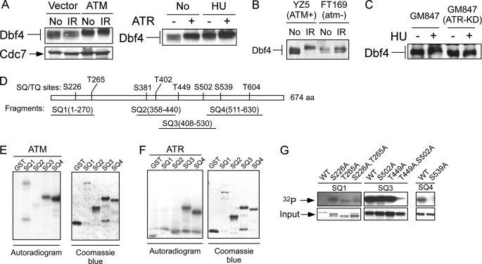
DNA damage-induced Dbf4 hyperphosphorylation is mediated by ATM and ATR. A, 293T cells were transfected with empty vector (Vector or −), the ATM expression vector (ATM, left) or the ATR expression vector (+, right). Whole cell extracts were prepared after mock treatment (No) or exposure to IR (20 Gy, 30 min after) or HU (1 mm, 24 h after), and Western blotting for Dbf4 was performed. B, whole cell extracts were isolated from ATM-deficient cell line (FT169; atm−) or its derivative cell line (YZ5; ATM+) reconstituted with wild type ATM before (No) or after exposure to IR (20 Gy, 30 min after). Western blot analysis for Dbf4 was performed. C, fibroblast cell line GM847 and its derivative cell line carrying doxycycline-inducible ATR-KD (GM847-ATR-KD) were incubated with doxycycline (1 μg/ml) for 24 h and then treated with HU (1 mm, 24 h, +) or without (−) in the presence of doxycycline. Whole cell extracts were prepared, and Western blot analysis of Dbf4 was performed. D, schematic drawing of full-length Dbf4 containing eight ATM/ATR consensus phosphorylation sites, (S/T)Q sites, is represented. Four GST-fused Dbf4 fragments (SQ1–SQ4) containing one or two (S/T)Q sites are illustrated. E, ATM in vitro kinase assay was performed by using anti-FLAG immunoprecipitates from 293T cells that were transiently transfected with FLAG-tagged ATM. The immunoprecipitates were incubated with each of the glutathione-eluted purified GST-SQ fragments as indicated in the presence of [γ-32P]ATP. The autoradiogram shows the results of the in vitro kinase assay (left), and the Coomassie Blue-stained gel indicates the relative amount of protein added into the in vitro kinase reaction (right). F, 293T cells were transiently transfected with FLAG-tagged ATR, and the anti-FLAG immunoprecipitates were incubated with each of the glutathione-eluted purified GST-SQ fragment in the presence of [γ-32P]ATP. Phosphorylation of Dbf4 fragments and the input of the Dbf4 fragments are shown by autoradiogram (left) or Coomassie Blue-stained gel (right), respectively. G, in vitro ATM kinase assay was performed using purified GST-Dbf4 wild type fragments SQ1, SQ3, or SQ4, or the corresponding fragments with alanine substitution of serine or threonine residues at the (S/T)Q (SQ/TQ) sites as indicated. ATM-mediated phosphorylation of Dbf4 fragments was revealed by an 32P autoradiogram (top), and relative input for the GST-fused Dbf4 fragments is indicated by a Coomassie Blue-stained gel (bottom).

