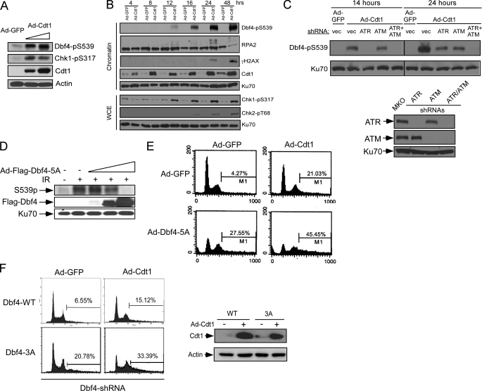
FIGURE 7.
ATM/ATR-dependent Dbf4 phosphorylation is important for suppressing DNA rereplication. A, whole cell lysates were prepared from U2OS cells infected with Ad-GFP or Ad-Cdt1 using two different viral titers for 48 h (5 × 10⁷ or 1 × 10⁸ pfu/ml). The lysates were analyzed by Western blotting using the indicated antibodies with actin as a loading control. B, U2OS cells were infected with Ad-GFP or Ad-Cdt1 (5 × 10⁷ pfu/ml), and whole cell lysates and chromatin fractions were prepared at different time points after infection. The prepared lysates and chromatin fractions were immunoblotted with the indicated antibodies, and Ku70 was used as a loading control. C, U2OS cells were retrovirally infected with vector pMKO (vec) or pMKO encoding shRNAs against ATR or ATM. These cells were subsequently infected with Ad-GFP or Ad-Cdt1 (5 × 10⁷ pfu/ml), and whole cell lysates were prepared at 14 or 24 h after infection. Western blot analysis was performed using the antibodies recognizing phosphorylated Ser-539 of Dbf4 or Ku70 (top). The expression of ATR or ATM was examined by Western blot analysis (bottom). D, U2OS cells were infected with Ad-GFP or Ad-Dbf4–5A (S226A/T265A/T449A/S502A/S539A) using increased viral titers (5 × 10⁷, 5 × 10⁸, or 1 × 10⁹ pfu/ml) for 48 h and subsequently treated with IR (20 Gy, 1 h after, +) or mock treatment (−). Western blot analysis was performed using antibodies specific for Dbf4-pS539 or FLAG tag. Ku70 was used as a loading control. E, U2OS cells were infected with Ad-GFP or Ad-Dbf4-5A (5 × 10⁸ pfu/ml) for 24 h, followed by infection using Ad-GFP or Ad-Cdt1 (5 × 10⁷ pfu/ml) for another 48 h. Infected cells were collected for FACS analysis. F, the expression of endogenous Dbf4 in U2OS cells expressing wild type Dbf4 or Dbf4-3A was suppressed by two rounds of shRNA retroviral infection, followed by adenoviral infection with Ad-GFP or Ad-Cdt1 (5 × 10⁷ pfu/ml). FACS analysis was performed 48 h after adenoviral infection (left). Cdt1 expression was shown by anti-Cdt1 Western blot analysis with actin as a loading control (right).

