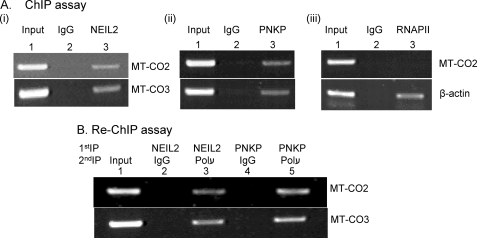
FIGURE 3.
A, ChIP assay. After sonication and immunoprecipitation of cross-linked chromatin separately with Ab for FLAG (panel i), PNKP (panel ii), or RNAP II (panel iii), the IPs were washed, the bound protein-DNA complexes were eluted, and the precipitated DNA was amplified by PCR using mitochondrial (MT-CO2 and MT-CO3) or nuclear (β-actin) gene-specific primers. B, re-ChIP assay. The bound fractions from the first IP were eluted, divided into two aliquots, and subjected to a second IP with IgG (as control) or with a specific Ab. PCR amplifications were performed using specific primers as shown in Table 1B.

