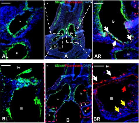
FIGURE 7.
Inhibition of hnRNPA2/B1 in NCCs results in developmental defects of 3rd and 4th branchial arch arteries. A and B, triple stainings for DAPI (blue), SMαA (green), and morpholino-oligonucleotides (red fluorescein) with sections from embryos electroporated with control (A) or Hnrnpa2/b1 (B) morpholino-oligonucleotides. In Hnrnpa2/b1 morpholino group, fluorescein-labeled NCCs could not migrate into branchial arch arteries at the right-hand side (BR, yellow arrows) and differentiate into SMCs (BR, white arrows), leading to the developmental defect of smooth muscle in the 3rd and 4th branchial arches (BR, indicated by red arrows). In contrast, the left-hand side of branchial arch arteries (BL) developed normally. In control morpholino group, both sides of branchial arch arteries showed complete smooth muscle formation (AR and AL). The fluorescein-labeled NCCs could not only migrate into branchial arch arteries wall at the right-hand side but also differentiate into SMCs in the 4th branchial arch artery. White arrows indicate those NCCs that are differentiating to SMCs. Representative images from five embryos in each group are presented here. Scale bar, 50 μm. Isotype IgG substituted primary antibody as negative control during staining process. iii, 3rd branchial arch artery; iv, 4th branchial arch artery. Original unmerged images of AR and BR were provided in supplemental Fig. S3.

