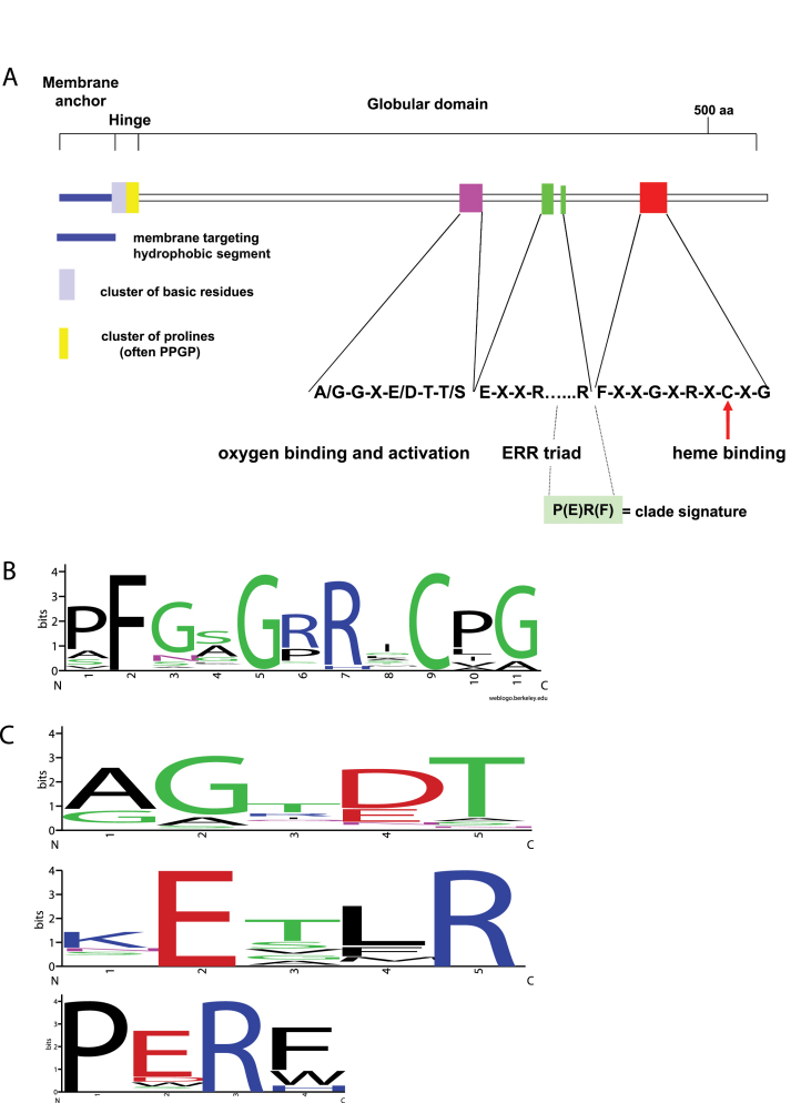
Figure 2.
Conserved structures and sequences in P450 proteins.
(A) Map of the signature motifs in the P450 proteins. The Glu and Arg of the K-helix consensus sequence (KETLR) and the Arg in the “PERF” consensus sequence form the E-R-R triad.
(B) WebLogo of the P450 heme binding motif constructed from A. thaliana P450s. The letter size is proportional to the degree of amino acid conservation. The F, G and C residues are conserved in all plant P450s. The C residue is universally conserved in all P450s across kingdoms and coordinates the iron in the heme.
(C) WebLogos of the other conserved structures and sequences in P450 proteins. AG×DT (I-helix), KETRL (K-helix) and PERF/W, respectively.

