Figure 42.
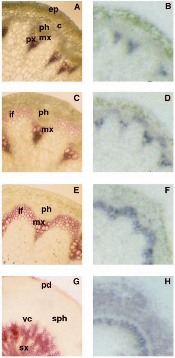
Immunolocalization of the expression of CYP98A3 in stems and roots.
Hand-cut transversal sections of inflorescence stems and roots were stained with phloroglucinol HCl, a red coloration reflecting lignin content. Adjacent sections were printed onto nitrocellulose and revealed using anti-CYP98A3 polyclonal antibodies. Blue staining is indicative of CYP98A3 expression. In stems, prints were taken at increasing distances from the apical meristem to monitor temporal and developmental expression of CYP98A3 in conjunction with the differentiation of lignified tissues. No blue staining was obtained with preimmune antibodies. (A,C,E), and (G), lignin staining with phloroglucinol; (B,D,F), and (H), immunostaining of CYP98A3. (A) and (B), upper segment of the stem, close to the flower bud; (C) and (D), mid-stem; (E) and (F), lower, well differentiated stem close to the rosette; (G) and (H), root, ep, epidermis; c, cortex; px, protoxylem; mx, metaxylem; ph, phloem; if, interfascicular region; sx, secondary xylem; vc, vascular cambium; sph, secondary phloem; pd, periderm. Reprinted from Schoch et al. (2001) with permission from American Society for Biochemistry and Molecular Biology.

