
Figure 50.
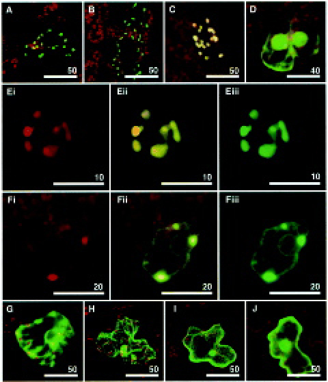
Confocal images of tobacco leaf cells following microprojectile bombardment showing chloroplast localization of CYP701 A3.
Images were taken 24 h after bombardment of tobacco leaves with tungsten particles coated with individual plasmid constructs. The cells expressing the GFP construct are epidermal cells with the exception of Ei-iii which show a mesophyll cell. The images are dual GFP (green) and chlorophyll (red) channels, except those labelled (i) and (iii) which show separate chlorophyll and GFP channels, respectively. The lengths of scale bars are given in µm in each panel. (A) TPCPS—GFP (copalyl diphosphate synthase transit peptide-GFP fusion: plastidial control), (B) TPKS—GFP (kaurene synthase transit peptide-GFP fusion: plastidial control), (C) RbcS—GFP, (D) smGFP, (E) TPCYP701A3—GFP, (F) CYP701A3—GFP, (G) TPCYP88A3—GFP, (H) TPCYP88A4—GFP, (I) mGFP5, (J) 20ox2—GFP Images reprinted from Heliwell et al. (2001) with permission from Wiley-Blackwell.