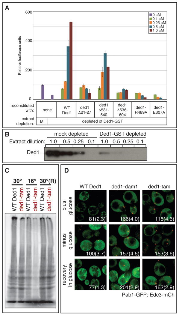
Figure 6. Ded1 ATPase and assembly domains promote translation.
A. Extracts that were mock or Ded1 depleted were used for in vitro translation reactions as in Figure 3A. B. Sequential dilution of mock and Ded1 depleted extracts analyzed by western blot with α-Ded1 antibody. C. yRP2799 containing either wild type DED1 or ded1-tam as the sole copy of DED1 was assayed for translation by 35S incorporation during growth at 30°C, after a 2 hour shift to 16°C, and after recovery for 1 hour at 30°C(R). D. yRP2799 containing either wild type DED1, ded1-dam1, or ded1-tam as the sole copy of DED1 were transformed with plasmid pRP1657, which encodes Edc3-mCh (a P-body marker) and Pab1-GFP (a SG marker). Strains were grown to mid-log phase and then shifted to 16°C for 2 hours. Localization was assessed in the presence of 2% glucose, after 15 minutes of glucose deprivation, and after a subsequent 15-minute recovery in 2% glucose. Each image includes quantitation of P-body intensity, normalized to wild type Ded1 after glucose deprivation, and the average number of P-bodies per cell, shown in parentheses. See also Figure S4.

