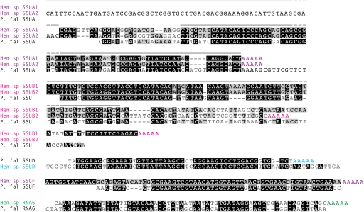
FIG. 3.—
Hematodinium sp. rRNA sequences aligned to those of their fragmented apicomplexan counterparts. Color groups indicate distinct rRNA fragments, with oligoadenylation also shown in color. Gray lines indicate positions of Northern blot probes. Hem. sp, Hematodinium sp.; P. fal, Plasmodium falciparum.

