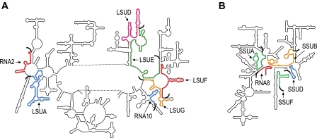
FIG. 4.—
Predicted locations of dinoflagellate LSU (A) and SSU (B) rRNA fragments, relative to Escherichia coli LSU and SSU secondary structures. The approximate locations of dinoflagellate rRNA fragment sequences are shown in color. Oligo(A) tails of dinoflagellate rRNA transcripts are represented as thick curved black lines.

