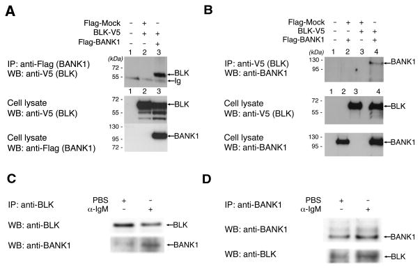
Figure 1. BANK1 and BLK Display a Protein-Protein Interaction.
a) Immunoprecipitation and western blot showing protein-protein binding of BANK1 and BLK. FLAG-BANK1 and BLK-V5 were co-transfected into HEK293T cells and immunoprecipitation was done using anti-FLAG antibodies. Western blot was performed using anti-V5 antibodies and confirmed with anti-FLAG antibodies. Lanes show: 1. Untransfected cells; 2. Co-transfection of FLAG-mock vector and BLK-V5; 3. Co-transfection of FLAG-BANK1 and BLK-V5.
b) Immunoprecipitation of cell extracts from co-tranfections showing recovery of BANK1 with the anti-V5 antibody directed to BLK-V5. Lanes show: 1. Untransfected cells; 2. Co-transfection of FLAG-Mock vector and FLAG-BANK1; 3. Co-transfection with FLAG-Mock and BLK-V5; and 4. Co-transfection of BLK-V5 and FLAG-BANK1.
c) Immunoprecipitation of endogenous BANK1 and BLK in the human cell line Daudi. Cell extracts were immunoprecipitated using anti-human BLK and the immunoprecipitates analyzed by Western blot.
d) Immunoprecipitation of endogenous BANK1 and BLK in naïve primary B cells. Cells were treated with anti-human IgM (SouthernBiotech) in a final concentration of 10 ug/ml for 10 minutes in serum-free RPMI medium or left unstimulated. Cell extracts were immunoprecipitated with anti-human BANK1 antibody (sc-133357, Santa Cruz Biotech) and analyzed by Western blot.

