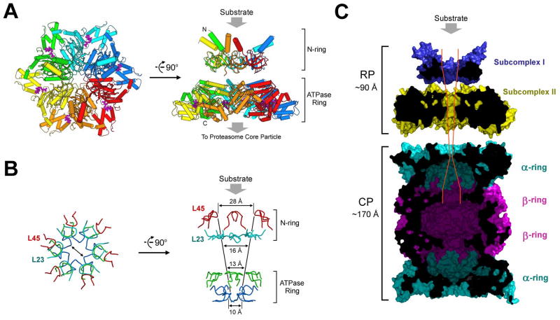
Figure 7.
Structural and functional implications on the proteasome. (A) A structure-based model of the PAN regulatory particle. Substrate protein is thought to bind to the distal face of subcomplex I as shown. (B) The constrictions are conferred by four layers of surface loops. The first two layers come from subcomplex I, involving loops L23 and L45, and the latter two layers come from the Ar-Φ and pore-2 loops in the nucleotidase domains. (C) A structural view of the complete proteasome in M. jannaschii. The modeled, complete proteasome is cut across the middle section to show the path and constrictions to the degradative chamber in the β-rings. The placement of subcomplexes I and II and their position relative to the 20S CP were based on measurement of the reported electron microscopic images of the PAN-20S complex (Smith et al., 2005).

