
Figure 1.
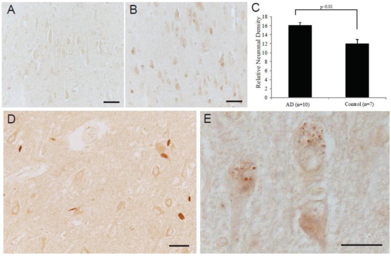
Localization of fluorophore immunoreactivity is increased in AD pyramidal neurons (B) as compared to an age-matched control (A). Densitometric analysis of the pyramidal neuron levels of fluorophore, shows significantly higher levels in the AD cases (n=10) compared to age-matched controls (n=7) (p<0.01, C). In the AD cases, many neurons contained intensely labeled GVD structures (B, and higher magnification in E) and some cases demonstrated labeling of Hirano bodies (D). Scale bar for A,B,D = 50 μm; E = 20 μm.