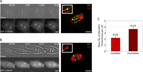
FIGURE 3:
Incomplete spindle pole separation at NEB promotes formation of merotelic kinetochore attachments. (A, B) Examples of GFP–γ-tubulin PtK1 cells imaged by time-lapse microscopy from NEB to late prometaphase, then immunostained for KTs and MTs, relocalized, and imaged by confocal microscopy (far right). A cell with complete spindle pole separation at NEB is shown in A, whereas a cell with incomplete spindle pole separation is shown in B. Insets represent 225% enlargements of the KTs indicated by the arrowheads. (C) The histogram shows that prometaphase cells with incomplete spindle pole separation at NEB exhibit higher (t test, p < 0.01) numbers of merotelic KTs than do cells with complete spindle pole separation at NEB. Error bars represent standard errors of the mean. Scale bars in the time-lapse images, 10 μm. Scale bars in the fixed cell images, 5 μm.

