FIGURE 1:
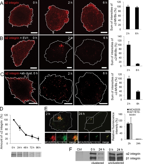
Clustered α2 integrin is degraded in α2-MVBs. (A) In SAOS-α2β1 cells, α2 integrin was labeled on the cell surface with anti–α2 integrin antibody (MCA2025) and unclustering goat anti–mouse Fab DyLight 549 fragment. The α2 integrin–Fab conjugate was then internalized for 0, 2, and 6 h and imaged with similar confocal settings. Confocal sections through the cells were projected together. Bars, 10 μm. (B) In addition to α2 integrin–Fab label (described in A), EV1 was bound to cells on ice before internalization. Cells were imaged as described. Bars, 10 μm. (C) α2 integrin was clustered with anti–α2 integrin MCA2025 and clustering goat anti–mouse Alexa 555 antibodies (+ab clust.). Intensity of fluorescence signal (A–C) was measured from confocal three-dimensional sections of single cells. Altogether 30 cells from three independent experiments were analyzed. Mean values ± SE are shown. (D) Normal turnover rate of α2 integrin was determined from metabolically labeled and immunoprecipitated samples. Quantification of gel bands was done with Adobe Photoshop, and the results are shown as averages of three independent experiments (±SE). (E) To evaluate the degradation of α2 integrin in α2-MVBs in more detail, integrin was labeled on the cell surface and after fixation again with another antibody: α2 integrin was first clustered with anti–α2 integrin MCA2025 antibody, followed by clustering with goat anti–mouse Alexa 555 antibody. After internalization for 2 and 24 h, cells were labeled with another antixα2 integrin antibody, biotinylated A211E10, and streptavidin–Alexa 488 (green). Fluorescence intensity was measured from confocal z-stacks of 30 cells from three independent experiments (±SE). Bars, 10 μm. (F) Degradation of α2 integrin was followed also after surface biotinylation of all proteins and by immunoprecipitating the integrin clusters (clustered) or unclustered α2 integrin (unclustered) via the clustering antibody or integrin antibody, respectively. Control cells were treated with the clustering secondary antibody without the primary antibody.

