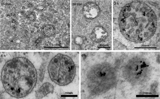
FIGURE 3:
EM images of endosomes triggered after α2β1 integrin clustering. Internalization for shorter time periods—for example, 15 min—shows structures that have tubular extensions and vesicular parts without clear ILVs. ILVs grow continuously during internalization, and after 30 min >45% of the structures show several ILVs. The majority of the structures after 2 h and later are MVBs with a high number of ILVs. After 6 h, the number of clearly defined ILVs seemed to have decreased, and for some structures the limiting membrane is also less conspicuous (lower right). Integrin was labeled on the plasma membrane with specific primary antibodies, followed by secondary antibodies and protein A gold (10 nm). Bars, 200 and 500 nm.

