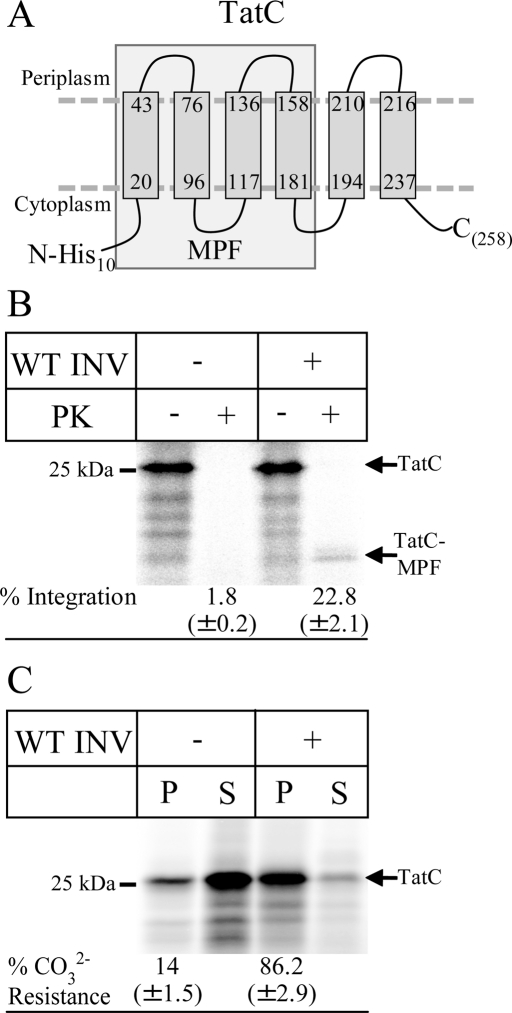
FIGURE 2:
Integration of in vitro–synthesized TatC into inner membrane vesicles. (A) Predicted topology of TatC according to Behrendt et al. (2004); a His10 tag was fused to its N-terminus. The boxed portion of TatC most likely corresponds to the 19-kDa, membrane-protected fragment of TatC (TatC-MPF). (B) 35S-Labeled His10-TatC was in vitro synthesized in the absence (–INV) or presence of wild-type E. coli inner membrane vesicles (WT INV; 2 mg/ml). After synthesis, one-fourth of the reaction was precipitated with TCA, and the remainder was first treated with 0.5 mg/ml PK for 30 min at 25°C and then TCA precipitated. Full-size TatC (TatC) and the TatC-MPF are indicated. Note that wild-type E. coli INV contains sufficient amounts of SRP and FtsY (Koch et al., 1999). The percentages of PK protection was calculated using ImageQuant (GE Healthcare) by quantifying the ratio of radioactivity present in the PK-treated sample and the directly TCA-precipitated sample and are the mean values of at least three independent experiments. Note that the calculation is corrected for the loss of methionine/cysteine residues and based on the assumption that TatC-MPF corresponds to the first four TMs of TatC. (C) TatC was in vitro synthesized as in B but extracted with alkaline Na2CO3 (pH 11.3, 0.2 M final concentration). After ultracentrifugation, pellet (P) and supernatant (S) were separated by SDS–PAGE. For quantification, the amounts of radioactive material in both fractions were set as 100%, and the distribution between both fractions was calculated. The values provided are the mean values of at least three independent experiments, and the SD is indicated.

