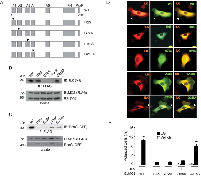
FIGURE 3:
RhoG/ELMO2/ILK complexes mediate EGF-induced keratinocyte polarization. (A) Schematic of the ELMO2 mutants used. Arrowheads indicate the position of mutated residues. A, ARM repeat; PH, pleckstrin homology domain; PxxP, proline-rich region. (B) Lysates from IMDF cells exogenously expressing mCherry/V5–tagged ILK together with FLAG-tagged ELMO2 proteins were used in immunoprecipitation assays with anti-FLAG antibodies. FLAG-ELMO2 immune complexes were resolved by SDS–PAGE and analyzed by immunoblot with anti-V5 antibodies to assess presence of ILK. The lysates were also analyzed by immunoblot with anti-FLAG or anti-V5 antibodies, as indicated. (C) Lysates from IMDF cells exogenously expressing GFP-tagged RhoG together with FLAG-tagged ELMO2 proteins were used in immunoprecipitation assays with anti-FLAG antibodies. FLAG-ELMO2 immune complexes were resolved by SDS–PAGE and analyzed by immunoblot with anti-GFP antibodies to assess presence of RhoG. The lysates were also analyzed by immunoblot with anti-FLAG or anti-GFP antibodies, as indicated. (D) Keratinocytes were transfected with vectors encoding mCherry/V5–tagged ILK and the indicated GFP-tagged ELMO2 protein. Twenty-four hours after transfection, the cells were briefly trypsinized, replated onto a laminin 332 substrate, and allowed to adhere for 3 h, prior to processing for direct fluorescence microscopy. Arrows indicate cell protrusions containing ILK and ELMO2. Bar, 20 μm. (E) Keratinocytes were transfected with vectors encoding the indicated proteins, as in D. Twenty-four hours after transfection, the cells were incubated in FBS- and EGF-free medium for 4 h, followed by stimulation with EGF or vehicle for 2 min. The cells were processed for fluorescence microscopy to assess the fraction of polarized keratinocytes. The results are expressed as the mean + SEM (n = 3). Where bars corresponding to vehicle-treated cells are absent, the fraction of polarized cells was <0.05%. *p < 0.05 relative to the corresponding vehicle-treated sample (ANOVA).

