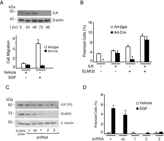
FIGURE 5:
Requirement for ILK and ELMO2 in EGF-induced polarization. (A) Ilkf/f keratinocytes were infected with adenovirus encoding Cre recombinase (Ad-Cre). Cell lysates were prepared at the indicated times after infection, resolved by SDS–PAGE, and analyzed by immunoblot with antibodies against endogenous ILK and β-actin (as loading control). Replicate cultures were infected with Ad-Cre or adenoviruses encoding β-galactosidase (Ad-βgal), and 72 h postinfection were trypsinized and used to measure EGF-induced chemotactic migration through Transwell inserts, as described in the Supplemental Materials and Methods. The fraction of keratinocytes that migrated through the inserts over 6 h was determined. The results are expressed relative to the fraction of cells infected with Ad-βgal that migrated in the absence of EGF, which is set to 1. The data represent the mean + SEM (n = 3). *p < 0.05 relative to Ad-βgal–infected cells stimulated with vehicle (ANOVA). (B) Ilkf/f keratinocytes were infected with Ad-Cre or Ad-βgal. Twenty-four hours after infection, cells were cotransfected with vectors encoding mCherry/V5–tagged ILK and/or GFP-tagged ELMO2. Twenty-four hours after transfection, cells were incubated in FBS- and EGF-free medium for 4 h, followed by stimulation with EGF for 2 min. The cells were processed for fluorescence microscopy, and the fraction of polarized cells was scored. Negative signs indicate cells that were transfected with vectors encoding only GFP- and/or mCherry. Results are expressed as the mean + SEM (n = 5). *p < 0.05 relative to cells infected with Ad-βgal (ANOVA). (C) Keratinocytes were transiently transfected with vectors encoding mCherry/V5–tagged ILK and either FLAG-tagged ELMO2 or with plasmids encoding control or ELMO2 shRNA sequences. Cell extracts were isolated 48 h after transfection, resolved by SDS–PAGE, and analyzed by immunoblot with antibodies against exogenous ILK, endogenous ELMO2 (except for the sample transfected with ELMO2-encoding vectors, which shows the total levels of exogenous pus endogenous proteins) or β-tubulin, used as protein loading control. sc, scrambled shRNA; v, vector only. The numbers indicate shRNA1, 2, or 3. (D) Cells transfected with shRNA-encoding vectors, as in C, were incubated in FBS- and EGF-free medium for 4 h, followed by stimulation with EGF for 2 min. The cells were processed for fluorescence microscopy, and the fraction of polarized cells was scored. Results are expressed as the mean + SEM (n = 3). Where bars are absent, the fraction of polarized cells was <0.05%. *p < 0.05 relative to cells treated with vehicle (ANOVA).

