
Figure 1. M1 assembles Fg into a cross-like pattern.
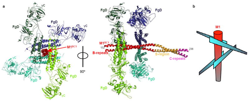
a, The M1BC1-FgD structure in ribbon representation. The M1BC1 B-repeats are in red, S-region in gold, and C-repeats in purple. The four FgD molecules bound by M1BC1 are in shades of blue or green, and the β2 integrin-binding γC domains are indicated. b, Schematic of the cross-like pattern of FgD (blue blades) surrounding M1BC1 (red cylinder).