Figure 1.

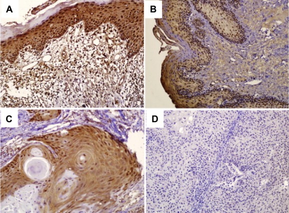
Immunohistochemistry analyses of MTUS1/ATIP expression in normal tongue, premalignant dysplasia and OTSCC tissue samples. Immunohistochemistry analyses for MTUS1/ATIP were performed as described in material and methods on A: normal tongue mucosa (n = 13), B: premalignant dysplasia (leukoplakia, n = 27), C: well differentiated primary SCC (n = 46), and D: moderately to poorly differentiated primary SCC (n = 34). Representative Images (×200) were shown.
