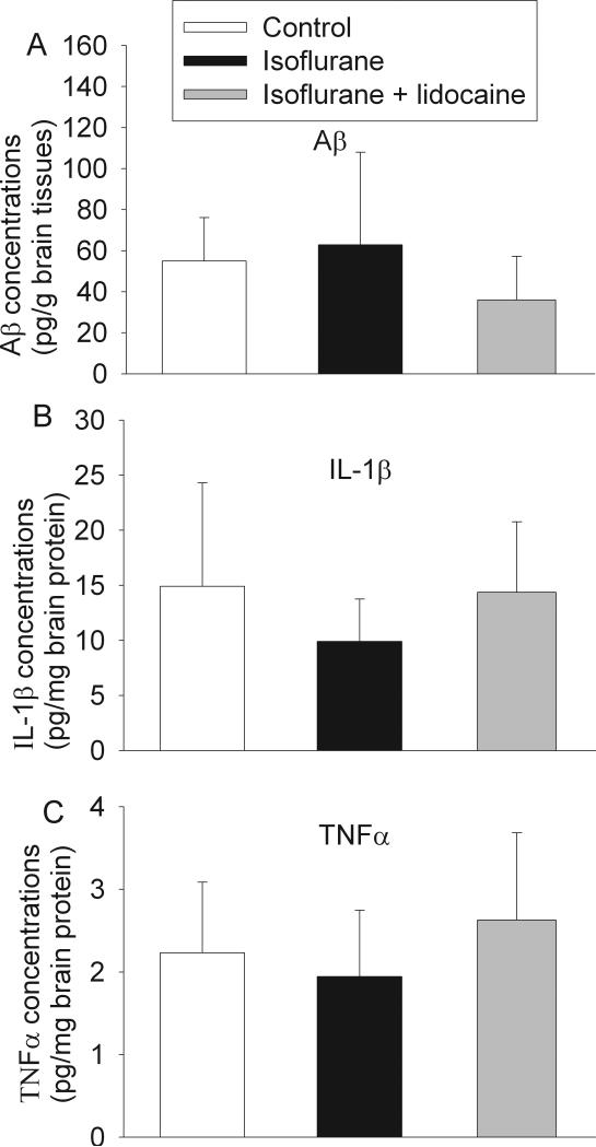
Fig. 4. Expression of β amyloid peptide (Aβ), interleukin (IL) 1β and tumor necrosis factor (TNF)-α in rat brains.
Eighteen-month-old Fisher 344 rats were exposed to or not exposed to 1.2% isoflurane in the presence or absence of lidocaine for 2 h. Aβ in the parietal cerebral cortex (panel A) and IL-1β (panel B) and TNFα (panel C) in the hippocampus were measured 29 days after the isoflurane exposure. Results are mean ± S.D. (n = 4 - 5).

