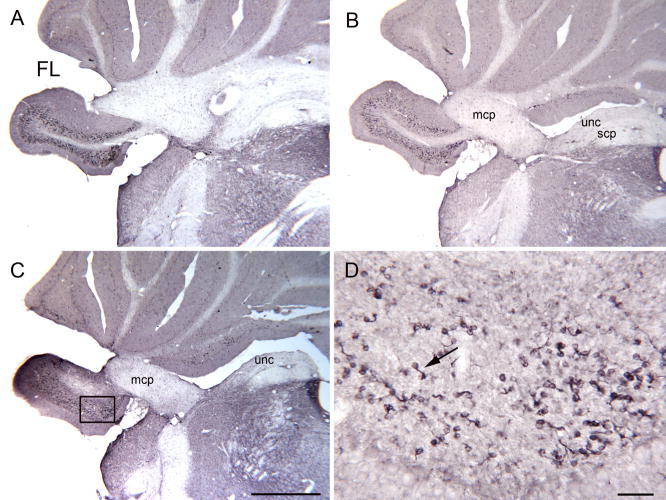
Figure 7.
A–C. DCX-ir UBCs in the rostral FL on three sections about 240 μm apart. C. The rectangle shows the location of the higher magnification image in D. Scale bar = 1 mm. D. Scattered DCX-ir UBCs (example at arrow). Scale bar= 50 μm. Abbreviations, scp, superior cerebellar peduncle; uc, uncinate tract

