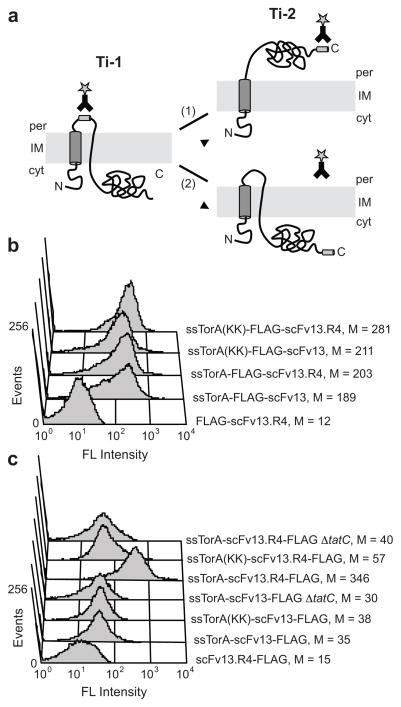
Figure 3. Separate detection of Ti-1 and Ti-2.
(a) Schematic of Tat translocation intermediates. Formation of Ti-1 can be detected when an epitope tag is inserted between the signal peptide and the N-terminus of the protein. If the protein is incorrectly folded (1), it cannot form Ti-2 and a C-terminal epitope tag is not accessible for immunolabeling. If the protein is properly folded (2), it is transported to the periplasm to form Ti-2, and a C-terminal epitope tag can be detected on the periplasmic face of the inner membrane. (b) FC analysis to detect Ti-1 for a poorly folded scFv (scFv13) and a well folded scFv (scFv13.R4). FLAG tags were inserted between the N-terminal Tat signal peptide and the scFvs. (c) FC analysis to detect Ti-2 for scFv13 and scFv13.R4. FLAG tags were placed at the C-terminus of scFvs. ssTorA(KK) indicates mutation of the Arg-Arg motif to Lys-Lys in the Tat signal peptide; ΔtatC indicates cells that lacked the TatC protein. FLAG tags were detected with a FITC-conjugated anti-FLAG antibody, and ssTorA-scFv13 lacking an epitope tag was included as a control. Median fluorescence values (M) are shown.

