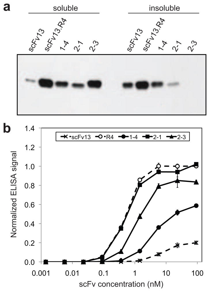
Figure 5. Expression and activity of scFv clones isolated using MAD-TRAP.
(a) Western blot analysis of soluble and insoluble fractions from cells expressing scFvs in the cytoplasm. Clone 1–4, 2-1, and 2–3 were isolated using MAD-TRAP. scFv13 was the starting sequence for the first-round library, and scFv13.R4 was isolated in a previous study after four rounds of directed evolution 22. Samples were normalized by total protein concentration in the soluble fraction, and blot was probed with an anti-6x-His antibody. (b) ELISA data for binding of isolated clones to βgal. scFvs were purified from cell lysate, and their binding to β-gal-coated ELISA plates was measured. Bound scFvs were detected with an anti-6x-His antibody. Data represent the average of six replicates and are normalized to the signal for scFv13.R4 at ~20 nM. Error bars represent standard error of the mean.

