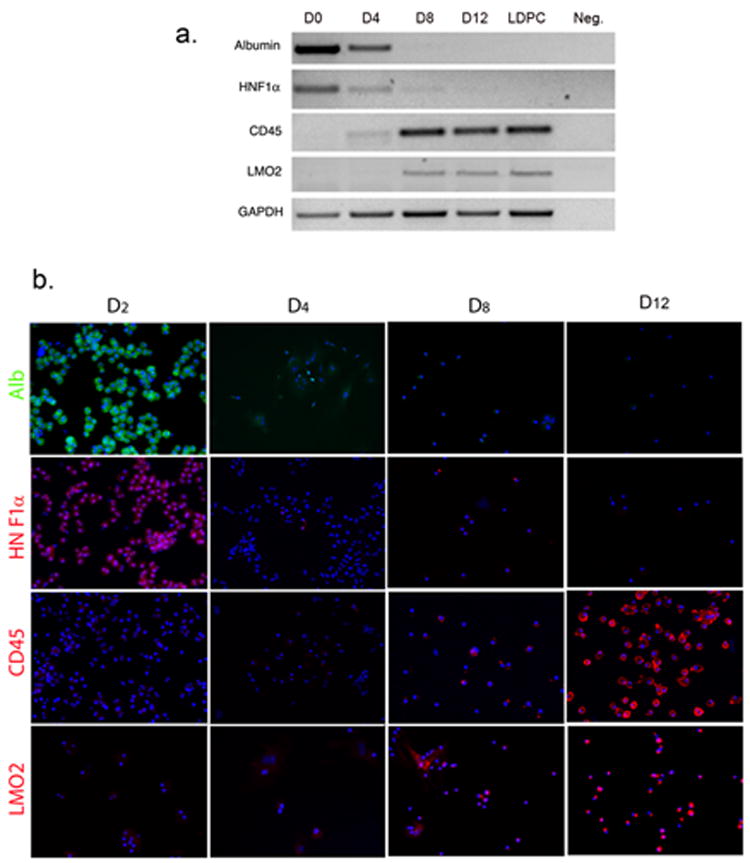
Figure 3.

Analysis of hepatocyte-specific and LDPC-specific markers at various time points during the LDPC culture period. (A) RT-PCR for the hepatocyte markers; albumin and HNF-1α, and LDPCs markers; LMO2, CD45. On day 0, only hepatocyte markers were expressed and no signals for LDPC markers were detectable. Beginning around day 4, hepatocyte markers became weaker and virtually gone by day 8 while LDPC markers showed an opposite trend and became progressively stronger. Day 12 cultures and “pure” LDPCs obtained on day 14 by gentle EDTA treatment of the cultures showed no difference indicating that the transformation into LDPCs was completed (only the cell number increased after day 12). The lane marked Neg. shows the negative control reaction. (B) IF studies of the LDPC cultures for the same markers confirmed the RT-PCR results again showing rapid disappearance of hepatocyte markers by day 4 and progressively stronger expression of LDPC markers after day 8 (cell nuclei stained by DAPI in blue, original magnification, 100x). The observed patterns were consistent with the rapid transformation of hepatocytes into LDPCs observed morphologically in Figure 2.
